LAB SESSIONS
Laboratory sessions will be available for a limited number of participants. The selection will be done in a first come first served basis.
Participants can select between the 4 following topics:
A. Microscopy image quantitative analysis using digital processing
B. Imaging Flow Cytometry, looking beyond the flow
C. Ultrasound imaging: development, physiology and disease
A. Microscopy image quantitative analysis using digital processing
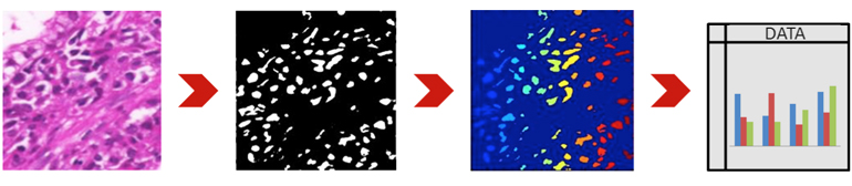
Digital image processing techniques enable fast and objective analysis of large amount of data. However, for succeeding in the data analysis, users must have adequate knowledge about the techniques. As there is no perfect solution for every cases of microscopy image data analysis, users must be familiarized with the wide range of choice of methodologies and their parameterization.
In this lab session we will focus on the digital image processing methodologies most relevant to microscopy image data analysis. Additionally, we will provide several standard approaches for analyzing data from the most usual image modalities. Strong emphasis will be given to the process of parameterization of such approaches and the problems that may arise from such analysis. Practical examples will be given and the participants are encouraged to provide examples from their own data.
B. Imaging Flow Cytometry, looking beyond the flow
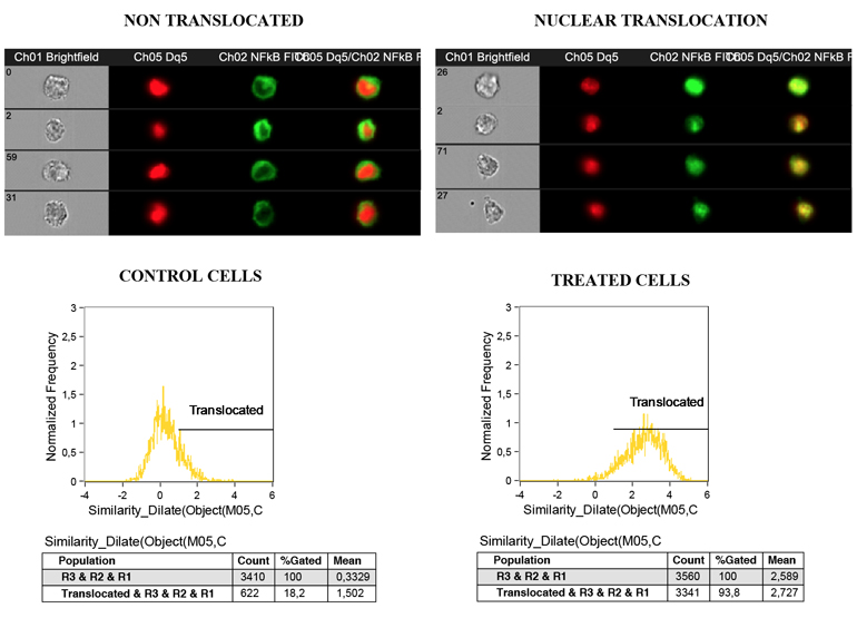
Imaging flow cytometry is a state of the art technology that combines the statistical strength, sensitivity, and high speed of a conventional flow cytometer by rapidly acquiring large number of cells with the quantitative image analysis of the detailed cellular imagery with morphologic (and functional) information captured by a microscope.
Similar to flow cytometry, fluorescence intensity and cell complexity can be analyzed, but furthermore and amongst others, morphological characterization, protein translocation and colocalization, can also be evaluated. The high versatility of the analysis software allows for the quantification of a diverse set of parameters granting the user complete autonomy in regard to the features to be included in the data analysis. From the choice of the feature to be analyzed, to the option of combining several features at the same time, or even the decision of the specific localization in the cell where the feature should be quantified, allowing the attainment of a multiplicity of results from one single experiment.
In this lab session, the students will learn how to use the imaging flow cytometer (ImageStreamX from Amnis) by running different samples, and to use the software IDEAS to analyze and quantify different parameters, such us probe internalization, spot counting, nuclear localization, organelle characterization and cell morphology.
C. Ultrasound imaging: development, physiology and disease
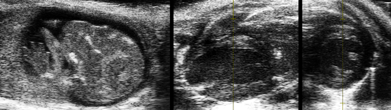
In small animal research, non-invasive analysis of diverse pathophysiological processes is of major importance. High resolution ultrasound imaging is particularly valuable for real-time evaluation of embryonic development, cardiac functional performance and cancer progression.
The course will comprise a hands-on laboratory session on non-invasive ultrasound mouse imaging using the high-frequency/high-resolution Vevo2100 platform. The students will monitor, by real-time ultrasound imaging, different setups:
(i) embryonic development,
(ii) cardiac performance under physiological and pathological conditions,
(iii) tumor growth and metastisation.
D. Confocal Raman spectroscopy and atomic force microscopy
Combining chemical analysis and nanoscale imaging in the study of cells and scaffolds
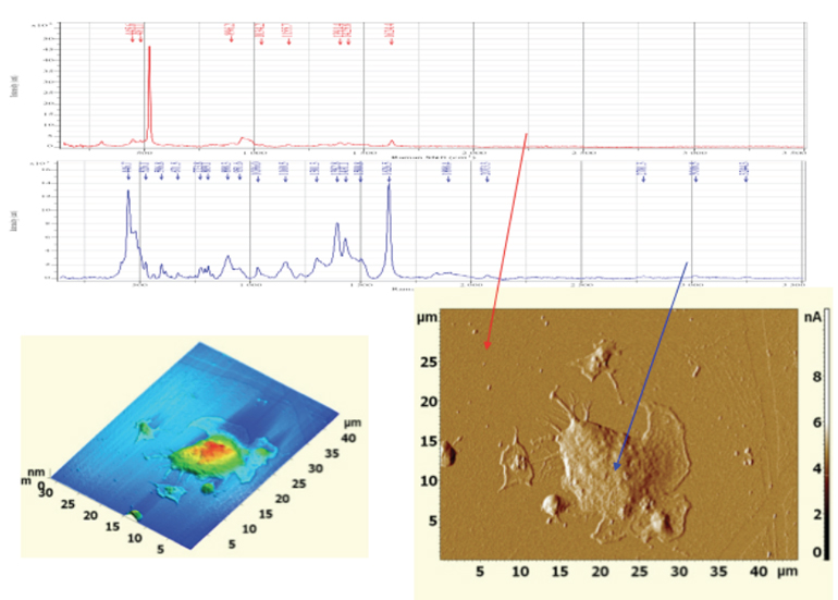
Confocal Raman spectroscopy probes the interaction of monochromatic laser light with chemical bonds, giving detailed insight into the chemical composition of a sample. On the other hand, Atomic Force Microscopy provides information on topographic and mechanical properties of a material, with molecular resolution. In this laboratory session the participants will have the opportunity to operate a system that combines both experimental techniques, in order to characterize cells and scaffolds.
| ORGANIZED BY: | CO-ORGANIZED BY: | SUPPORTED BY: | |
| Rua do Campo Alegre, 823 4150-180 Porto - Portugal Tel +351 226 074 900 Email: bioimaging2014@ineb.up.pt |
  |
 |